Introduction

The membrane’s ability to control the transport of materials across the membrane is one of the essential life processes in organisms. The membrane determines what solutes shall enter or leave the cell, which involves complex structures functioning in the transfer of molecules across the membrane. And any mutation or malfunction in this transport mechanism leads to severe life-threatening diseases.

The transport of essential molecules is a series of reactions regulated by the complex interaction between the macromolecules that make up the membrane. These macromolecules are proteins, lipids, and carbohydrates.

The biological membrane is semipermeable, meaning it allows the transport of only a few molecules, ions, and water across the membrane. And its permeability for the molecules varies in different organisms based on the structural pattern of the lipid bilayer.[1]

The bilayer constitutes the membrane’s hydrophobic core, approximately 40 Å (4 nm) thick.[1] The integrated proteins spanning the membrane form the carrier and channel transport vehicle involved in catalyzing the translocation of solutes across biological membranes. These solutes include ions, nutrients, neurotransmitters, and numerous drugs.[2]

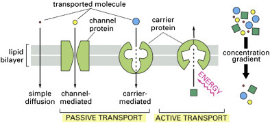
The transport of these essential biological molecules across the membrane is classified into two categories:

    • Passive Transport: It’s the transport mechanism of substances down the concentration gradient and without energy expenditure. It includes diffusion and facilitated diffusion.
    • Active Transport: It’s the transport of molecules across the membrane against the concentration gradient but with energy expenditure.
Basic types of membrane transport

Image: An illustration of the basic types of membrane transport: simple passive diffusion, facilitated diffusion (by channels and carriers), and active transport.

Credit: Stillwell W. (2016).[1]

Any mutation in molecules building the membrane transporters or involved in the transport mechanism can cause severe diseases like autism, epilepsy, migraine, depression, drug abuse, and cystic fibrosis.[2]

In this article, we’ll learn in-depth about the active transport of molecules across the membrane, their types, and a few examples of active transport in different organisms.

What Is Active Transport?

Active transport is a transport mechanism of molecules across the membrane from a region of lower concentration to one of higher concentration (against the concentration gradient) by utilizing energy (often ATP). Sometimes, an electrochemical gradient drives the transport.

Some examples of active transport include the uptake of glucose in the intestine of the human body and the uptake of minerals or ions into the root hair cells of plants.[3]

Types of Active Transport

Active transport is classified into two based on how energy is coupled to fuel the transport mechanisms. The classes are primary and secondary active transport.

1. Primary Active Transport

Primary active transport is also known as direct active or uniport transport. In this form of transport, molecules are transported across the membrane by breaking down adenosine triphosphate (ATP).

Molecules transported through this mechanism include ions such as Na+, K+, Mg2+, and Ca2+. Ion pumps or ion channels facilitate the transport of these charged molecules throughout the body.[4]

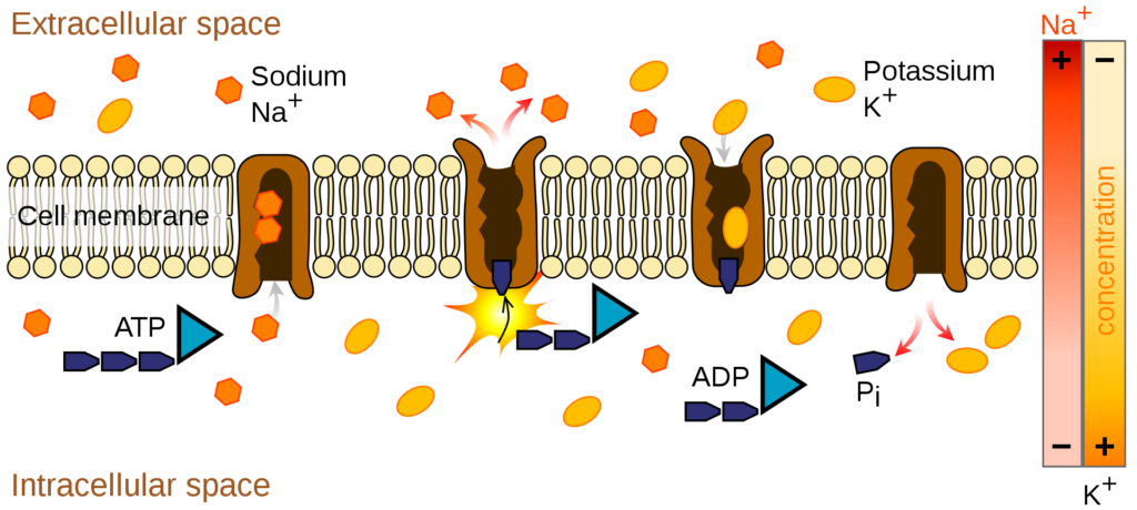
Example of primary active transport across the membrane

Image: The illustration of an example of primary active transport across the membrane.

Credit: Wikipedia[4]

Types of Primary Transport

1. P-type ATPase

It is also known as E1-E2 ATPases because of their ability to interconvert between two conformations (E1 and E2). They are ion and lipids pumps found in bacteria, archaea, and eukaryotes.[5]

The “P-type” denotes their ability to autophosphorylate the components within the pump, like aspartate and ATP. Structurally, they contain four domains responsible for their functions, which include the Phosphorylation (P) domain, Nucleotide-binding (N) domain, Actuator (A) domain, and Regulatory (R) domain.[5]

Examples include the sodium-potassium pump (Na+/K+-ATPase), calcium pump (Ca2+-ATPase, proton-potassium pump (H+/K+-ATPase), and proton pump (H+-ATPase) of plants and fungi.[5]

For better understanding, let’s have a look at the mechanism of the sodium-potassium pump (Na+/K+-ATPase):[6]

    • The carrier protein opens to the cell interior, allowing sodium ions to adhere to the high-affinity pump.[6]
    • When sodium binds to the carrier protein, it induces the phosphorylation of the pump via ATP hydrolysis.
    • The chemical modification to the pump due to phosphorylation causes it to undergo a conformational change. This change allows the pump to lose its affinity towards sodium, thus, releasing the sodium ions to the cell exterior or extracellular area.[6]
    • Though the conformational change in the pump causes sodium to lose its affinity, it creates a high-affinity environment for potassium ions on the pump. Thus, potassium ions bind to it and release the attached phosphate group.[6]
    • The phosphate released induces the pump to reassume its earlier confirmation, causing it to re-open inside the cell.
    • This new change in conformation changes the affinity of the pump from potassium to sodium ions. And the cycle continuously repeats itself.
    • Ouabain, a cardiac glycoside, is the inhibitor of sodium-potassium ATPase. It binds to the surface of the pump, and inhibits its dephosphorylation, thus, blocking K+ transport into the cell.[6]
Mechanism of the Na+, K+-ATPase

Image: Mechanism of the Na+, K+-ATPase.

Credit: Stillwell W. (2016).[1]

In humans, these pumps are involved in diverse functions, including nerve impulses, relaxation of muscles, secretion and absorption in the kidney, absorption of nutrients in the intestine, and other physiological processes.[1]
2. F-ATPase

It is also known as ATP synthase or ATP phosphohydrolase (H+-transporting).[7] These ATPases/synthases are found in mitochondrial inner membranes (in oxidative phosphorylation as Complex V) and chloroplast thylakoid membranes.[7]

It drives ATP synthesis by allowing the passive flux of protons across the membrane down their electrochemical gradient. The energy created during the transfer of protons releases the newly formed ATP from the active site of the F-ATPase.

But under low driving force conditions, these ATP synthases function as ATPases, generating a transmembrane ion gradient at the expense of ATP hydrolysis.

Structurally, it’s composed of two domains:

  • Fo Domain: It is integrated into the membrane and composed of three integral proteins classified as a, b and c. It’s responsible for ion translocation across the membrane.[7]
  • F1 Domain: It is composed of 5 polypeptide units – α (in 3 copies), β (in 3 copies), γ, δ, and ε. It carries the catalytic sites for ATP synthesis and hydrolysis.[7]

Examples of this transport system include mitochondrial ATP synthase and chloroplast ATP synthase.

3. V-ATPase

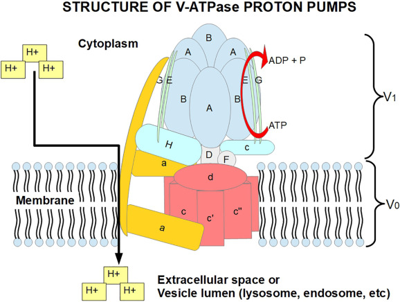
The vacuolar proton-translocating ATPases are ATP-driven pumps located in the tonoplast of the cell. The enzyme couples the energy from ATP hydrolysis to transport protons across intracellular and plasma membranes of eukaryotic cells. This function makes multiple cellular organelles more acidic than the surrounding cytoplasm.[8]

Any mutational changes in the enzyme can cause several implications, including cancer, neurodegenerative disease, and aging.[8]

Structurally, V-ATPases are complex multimeric proteins with two functionally separate domains. Both domains also have multiple tissue-specific isoforms present in different organisms:[8]

    • Domain V1: It is composed of eight subunits that perform ATP hydrolysis.[8]
    • Domain Vo: It consists of five subunits that are in charge of H+ transport through the membrane.[8]
Structure of V-ATPase proton pumps

Image: An illustrative structure of V-ATPase proton pumps.

Credit: Wikipedia[4]

4. ABC (ATP binding cassette) transporter

ABC transporters are an extremely diverse class of transporter proteins. It couples energy obtained through ATP hydrolysis to the movement of solutes across biological membranes. It is mainly identified in organisms belonging to the three major domains: bacteria, archaea, and eukarya.[9]

Structurally, it’s composed of four parts: two membrane-integral domains, each of which spans the membrane six times, and two ATP-hydrolyzing domains (or ABC subunits/domains).[9]

In humans, around 48 ABC genes are reported, among which most of them are related to diseases like cystic fibrosis, adrenoleukodystrophy, Stargardt disease, drug-resistant tumors, Dubin–Johnson syndrome, Byler’s disease, progressive familial intrahepatic cholestasis, X-linked sideroblastic anemia, ataxia, and persistent and hyperinsulinemic hypoglycemia.[10]

The members of this family are involved in several processes such as signal transduction, protein secretion, drug and antibiotic resistance, antigen presentation, bacterial pathogenesis, and sporulation. Its examples include Multidrug Resistance Transporter (MDR) and Cystic fibrosis transmembrane conductance regulator (CFTR).[9]

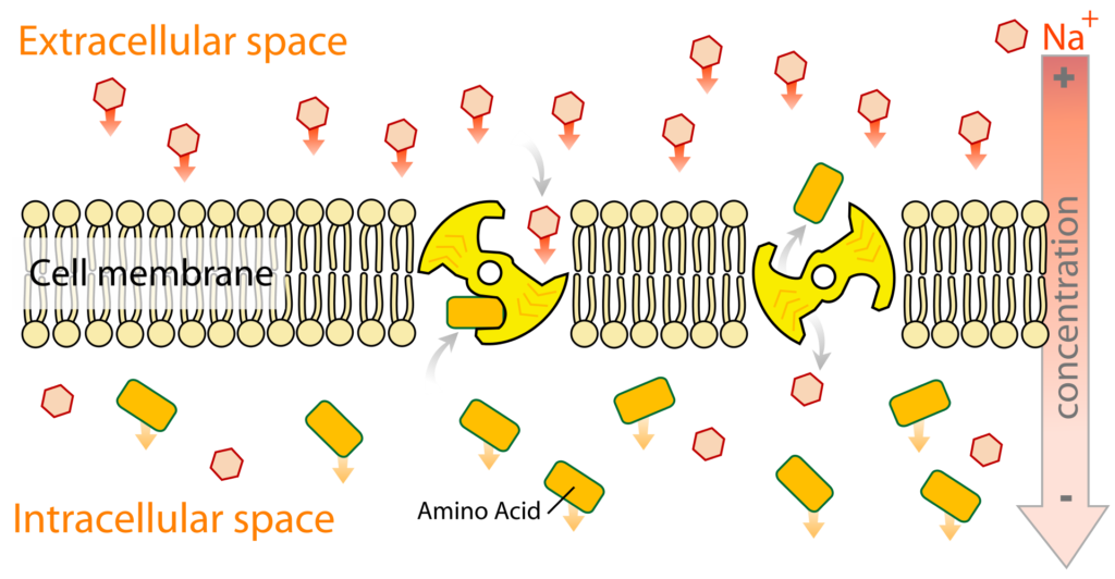
2. Secondary Active Transport

Secondary active transport is also known as coupled transport or cotransport, and it has two separate functions:[1]

    • 1st: The ion’s electrochemical gradient is generated across the membrane due to the energy-dependent movement of an ion (e.g., H+, Na+, or K+).[1]
    • 2nd: The generated ion gradient above is coupled to a solute’s movement in either the same direction (symport) or opposite direction (antiport).[1]

The transport of the first solute or ion (also known as driving ion) is through facilitated diffusion, whose sole purpose is to drive the transfer of the second solute across the membrane against its gradient.[1]

Secondary active transport across the membrane

Image: An illustration of secondary active transport across the membrane.

Credit: Wikipedia[4]

Types of Secondary Active Transport

Based on the transfer direction of two molecules/solutes across the membrane, the active transport is classified into two groups: Antiport and Symport.

For both antiport and symport, the concentration capacity of the transport process measures the level of effective transport.[11] The concentration capacity is defined as how well the driven ion/molecule is concentrated against a concentration gradient.[11]

For example, in Na+/glucose cotransporter, the ratio of the concentration of glucose in and out of the membrane, ([Glucose]i/[Glucose]o), decides the capacity of the transporter in concentrating glucose inside the cell against a concentration gradient.[11]

The concentration capacity is directly proportional to the ion/substrate coupling stoichiometry of transporters per transport cycle.[11]

For example, the coupling stoichiometry of the intestinal Na+/glucose cotransporter is two Na+ ions to one glucose molecule (2:1) per transport cycle. The higher the ion/substrate coupling ratio, the higher the concentration capacity of the transporter.[11]

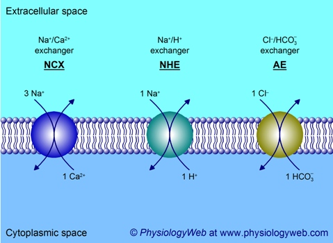
  • Antiport: In this system, two ions or other solutes are pumped in opposite directions across a membrane. It is also known as an exchanger or counter-transporter.[11] Examples are Na+/Ca2+ exchanger (NCX), Na+/H+ exchanger (NHE), and Cl/bicarbonate exchanger.
    • Na+/Ca2+ exchanger (NCX): It is ubiquitously found mainly in many cells and tissues of organisms and has an essential role in cytoplasmic Ca2+ homeostasis. This transporter transports three Na+ ions down the Na+ electrochemical gradient and one Ca2+ against its electrochemical gradient.[11]
    • Na+/H+ exchanger (NHE): It’s also a ubiquitously found anti-transporter that has an important function in regulating cytoplasmic pH. One Na+ is transported down the electrochemical gradient and one H+ against the gradient with 1:1 coupling stoichiometry.[11]
    • Cl/bicarbonate exchanger: It’s an electroneutral exchanger as it transports one Cl down the electrochemical gradient and one HCO3- against its electrochemical gradient.[11]
Three examples of antiport transport

Image: An illustration of the three examples of antiport transport.

Credit: Physiologyweb[11]
  • Symport: In this system, two molecules are transported in the same direction. One molecule is transported downhill, from high to low concentration, and another molecule moves uphill, that is, from low to high concentration (against its concentration gradient).[11] Examples of symporters are Na+/glucose cotransporter (SGLT1), GABA transporter (GAT), H+/oligopeptide transporter (PepT), and Sodium-coupled bicarbonate cotransporters (NBC).[11]
      • SGLT1 (sodium-glucose transport protein-1): It is present in the intestinal epithelium. Here, two Na+ is transported down the concentration gradient to transport one glucose molecule into the bloodstream across the apical membrane against its gradient.
        Such transport that leads to a net translocation of charge across the membrane is said to be electrogenic. Whereas it’s called the electroneutral process when no net charge is transported across the membrane per transport cycle.[11]
      • GABA transporter (GAT): It belongs to a large family of Na+ and Cl-coupled transporters. It regulates the basal concentration of the inhibitory neurotransmitter γ-aminobutyric acid (GABA) in the nervous system, and additionally, it regulates its concentration and lifespan in the synaptic cleft.[11]
      • H+/oligopeptide transporter (PepT): It is located in the apical membrane of the small intestine epithelial cells and proximal renal tubules. Here, one H+ is transported down the concentration gradient by transporting one dipeptide or tripeptide across the membrane, in the same direction, against the concentration gradient.[11]
        Further, it also serves as the entry route for peptidomimetic drugs such as β-lactam antibiotics. It has a dominant role in nitrogen absorption in the small intestine and reabsorption in the kidney tubules.[11]
      • Sodium-coupled bicarbonate cotransporters (NBC): It belongs to the family of Cl/bicarbonate exchangers and has an important role in acid-base balance in the body.[11]
        The basolateral membrane of proximal kidney tubules mediates the outward translocation of one Na+ ion and three HCO3- ions into the interstitial fluid.
Other than primary and secondary active transports, two other processes involving the transfer of molecules against the concentration gradient with energy expenditure include bacterial lactose transport and bulk transport.[11]

3. Bacterial Lactose Transport

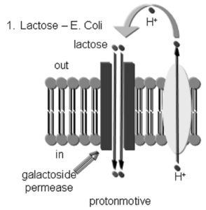
Bacterial lactose transport works similarly to secondary active transport.[1] The proton (H+) gradient generated by the bacterial electron transport system drives the lactose uptake (glucose-4-𝛃-D-galactoside) in bacteria. It’s essential to note that taking up lactose is a combination of the electrical gradient and pH gradient.[1]

The transport is performed by lactose permease, a membrane protein that is a member of the major facilitator superfamily. The proteins span the membrane with 12 transmembrane alpha-helices and have a molecular weight of 45,000 Daltons.[1]

Active transport of lactose in E. coli
Image: An illustration of active transport of lactose in E. coli.

Credit: Stillwell W. (2016).[1]

4. Bulk Transport

Bulk transport includes endocytosis and exocytosis that uses energy to transport materials across cells via vesicles.

  • Endocytosis: In this case, the cell membrane folds around or invaginates, forming a pocket around the material to be transported.[12] Its types include:
    • Phagocytosis: It’s the process of engulfing solid particles by the cell. Examples include a defense mechanism performed by white blood cells against bacteria or any other pathogen. The neutrophils surround, engulf, and destroy the microorganisms invading the human body.[12]
    • Pinocytosis: It’s the process of engulfing liquid particles by the cell.
    • Receptor-mediated endocytosis: It’s the targeted variation of endocytosis. The receptors present on the membrane bind to the specific particle or protein molecule, and the plasma membrane invaginates, bringing the substance and proteins into the cell.[2] The low-density lipoproteins (LDL) are removed from the blood by receptor-mediated endocytosis.
Different types of endocytosis
Image: An illustration of different types of endocytosis.

Credit: Lumenlearning[12]
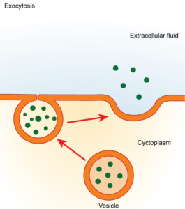
  • Exocytosis: It’s the transport of molecules to the outside of the cell, in the extracellular fluid of the organisms. It’s facilitated by the fusion of a vesicle with the plasma membrane that ensures the delivery of the particular molecule at the target location.[12]
transport of molecules into the extracellular fluid by vesicular fusion with the membrane
Image: An illustration of the transport of molecules into the extracellular fluid by vesicular fusion with the membrane.

Credit: Lumenlearning[12]

Conclusion

Membrane transport in organisms facilitates the transport of essential biomolecules and ions throughout the body thus, essential for organisms to thrive. Several types of transport are present in organisms, and only active transport is involved in the transport of molecules against the concentration gradient at the expense of energy. In this process, the transport of materials is aided by carrier proteins.

Active transport is primarily of two types, primary and secondary active transport. The primary active transport is uniport, whereas, in the secondary active transport, the uphill transfer of molecules across the membrane is coupled through the transfer of an ion or other molecule down the gradient.

Some examples of membrane proteins or transporters that facilitate the transport of molecules include sodium-potassium pumps, ABC transporter, and sodium-coupled bicarbonate cotransporters (NBC).

Any mutational changes in these transporters can cause life-threatening diseases. Scientists are diving deep into the functioning of these transporters to understand their mechanism and use with respect to clinical significance.

References

  1. Stillwell, W. (2016). Membrane Transport. An Introduction to Biological Membranes, 423–451. doi:10.1016/b978-0-444-63772-7.00019-1.
  2. Quick, M., & Javitch, J. A. (2007). Monitoring the function of membrane transport proteins in detergent-solubilized form. Proceedings of the National Academy of Sciences, 104(9), 3603–3608. doi:10.1073/pnas.0609573104.
  3. Active transport. Retrieved from https://byjus.com/biology/active-transport/
  4. Active transport. Retrieved from https://en.wikipedia.org/wiki/Active_transport
  5. P-type ATPase. Retrieved from https://en.wikipedia.org/wiki/P-type_ATPase
  6. Chen I, Lui F. Physiology, Active Transport. [Updated 2020 Sep 13]. In: StatPearls [Internet]. Treasure Island (FL): StatPearls Publishing; 2021 Jan-. Available from: https://www.ncbi.nlm.nih.gov/books/NBK547718/.
  7. F-ATPase. Retrieved from https://en.wikipedia.org/wiki/F-ATPase
  8. Koltai, T., Reshkin, S. J., & Harguindey, S. (2020). The vacuolar H+ ATPase proton pump. An Innovative Approach to Understanding and Treating Cancer: Targeting pH, 177–191. doi:10.1016/b978-0-12-819059-3.00008-3
  9. Erwin Schneider, Sabine Hunke, ATP-binding-cassette (ABC) transport systems: Functional and structural aspects of the ATP-hydrolyzing subunits/domains, FEMS Microbiology Reviews, Volume 22, Issue 1, April 1998, Pages 1–20, https://doi.org/10.1111/j.1574-6976.1998.tb00358.x.
  10. ATP-binding cassette transporter. Retrieved from https://en.wikipedia.org/wiki/ATP-binding_cassette_transporter
  11. Secondary Active Transport. Retrieved from https://www.physiologyweb.com/lecture_notes/membrane_transport/secondary_active_transport.html.
  12. Active Transport. Retrieved from https://courses.lumenlearning.com/suny-biology1/chapter/active-transport.